 University Journal of Surgery and Surgical Specialities
AN UNUSUAL PRESENTATION OF SCHWANNOMA NECK
Abstract
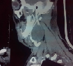
62 yr old male patient presented with (R) sided painless neck swelling for past 5 months with no pressure symptoms. On examination 6x5 cm swelling present in upper part of (R) side of neck which is well defined, mobile, variable in consistency. Examination of lower cranial nerves and oral cavity examination was normal. Provisional diagnosis of secondaries neck (R). But panendoscopy was normal. CT Neck showed possibility of metastatic node in (R) carotid space.1st FNAC showed carcinomatous deposits from thyroid but thyroid FNAC was normal. Hence Trucut biopsy taken from node shows spindle cell tumour with possibility of fibromatosis . So planned for excision biopsy. Intraoperatively tumor was wedged between ICA and ECA and found to be arising from nerve sheath of vagus nerve. Post op uneventful. HPE showed Schwannoma
Full Text:
PDFReferences
Som P, Sacher M, Stollman A, Biller H, Lawson W. Common tumors of the parapharyngeal space: refined imaging diagnosis. Radiology. 1988;169:81–8
Som PM, Braun IF, Shapiro MD, Reede DL, Curtin HD, Zimmerman RA. Tumors of the parapharyngeal space and upper neck: MR imaging characteristics. Radiology.1987;164:823–829
Furukawa M, Furukawa MK, Katoh K, Tsukuda M. Differentiation between schwannoma of the vagus nerve and schwannoma of the cervical sympathetic chain by imaging diagnosis. Laryngoscope. 1996;106:1548–1552. doi: 10.1097/00005537-199612000-00021
Refbacks
- There are currently no refbacks.

This work is licensed under a Creative Commons Attribution-NoDerivatives 4.0 International License.
An Initiative of The Tamil Nadu Dr MGR Medical University