Angioleiomyoma Uterus,Unique Variant of Uterine Leiomyoma
Abstract
Abstract
Uterine leiomyomas or fibroids are benign smooth muscle tumors of the uterus originates from smooth muscle and has thick blood walled vessles. Most women are asymptomatic while others complain of painful or heavy period and abdominal mass. Uterine angiolieomyoma is one of the extremely rare variant and only15 cases have been reported in the literature up to date. The case described here is about a 48 year old women who presented with abdominal distension and pain.
Introduction
Uterine angioleiomyoma is an extremely rare and unique variant of leiomyoma otherwise known as vascular leiomyoma originating from smooth muscle cells and containing thick-walled vessels and usually occurs in subcutaneous tissue, most often in the lower extremities and very rarely in the uterus.1,2 They occur middle-aged women and can present as an abdominal mass or with symptoms of abdominal pain and menorrhagia.3 These tumors can undergo spontaneous rupture and cause catastrophic intra-abdominal bleeding. A diagnosis of AL was made on histopathologic examination.
Case report
A 48 year old postmenopausal woman, came to the out patient department of gynaecology at SMIMS, Kulasekharam with complaints of lower abdominal pain for 1 month which was intermittent, dull aching and abdominal distension. She had attained menopause at the age of 45 years. Her obstetric score was P2l2A1 sterilized with all being full term normal deliveries.
On examination, she is moderately built, BMI – 27.40 kg/sq.m2, No pallor,lymphadenopathy, and oedema. Her vitals were stable and systemic examination showed no abnormalities.Per abdomen she was found to have a 22 weeks size uterus, non tender mobility restricted. On local examination, vulva and vagina shows atrophic changes, but per speculum examination showed a cervix with posterior lip hypertrophy, no discharge.
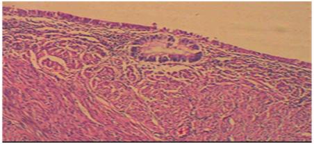
Per vaginal examination showed a 22 weeks uterus, non tender, firm in consistency, mobile and bilateral for nix freePost operative period was uneventful. Histopathology report came as a Angioleiomyoma uterus.On investigations she was found to have a haemoglobin level of 12 g/dl.USG showed a uterus of size (11.5 x 5.3 x 5.2 cm), with hypoechoic mass, lesion arising from posterior myometrium of uterus (15x11cm) is seen extending upto to level of umbilicus. Moderate vasularity seen. A subserous fibroid (3.5x2.2cm) is seen arising from posterior myometrium of lower body.Endometrium not separately visualized.Ovaries not visualised. PAP smear showed negative for intraepithelial lesion.She underwent Total Abdominal Hysterectomy with bilateral salpingo-oophorectomy 9/3/17.Intraoperatively she was found to have uterus enlarged in size 26-28 weeks variable in consistency, highly vascular,both ovaries&tubes.Post operative period was uneventful. Histopathology report came as a Angioleiomyoma uterus.
Full Text:
PDFReferences
Sharma C, Sharma M, Chander B, Soni A, Soni PK. Angioleiomyoma uterus in an adolescent girl: a highly unusual presentation.J.Pediatr Adolesc Gynecol. 2014;27:e69–e71.
Hachisuga T,Hashimoto H, Enjoji M. Angioleiomyoma.A clinicopathologic reappraisal of 562 cases. Cancer 1984;54:126-130
GargG,Mohanty SK. Uterine angioleiomyoma:a rare variant of uterine leiomyoma.Arch Pathol Lab Med 2014;138(8):1115-8
ManimekhalaP,KumarS,RamaniD,KrishnaReddy M..C.H,Ratna kumara.V.Uterine angioleiomyoma:A report of 2 cases. J Evol Med Dent Sci 2013; 2(18):3133-6.
Thomas S, Radhakrishnan L, Abraham L, Matthai A. Uterine angioleiomyoma with atypia,raised CA-125 levels, and Pseudo-Meigs syndrome: An Alarming Presentation.Case reports in Pathology 2012;(2012),Article ID 519473
Zizi-Sermpetzoglou A, Myoteri D, Arkoumani E, Koulia K, Tsavari A, Alamanou E, et al. Angioleiomyoma of the uterus: report of a distinctive benign leiomyoma variant. Eur J Gynaecol Oncol 2015;36:210-12.
Keerthi R, Nanjappa M, Deora SS, Kumaraswamy SV. Angioleiomyoma of cheek: report of two cases. J Maxillofac Oral Surg 2009;8:298-300.
Ishikawa S, Fuyama S, Kobayashi T, Taira Y, Sugano A, Iino M.Angioleiomyoma of the tongue: a case report and review of the literature. Odontology 2016;104:119-22.
Refbacks
- There are currently no refbacks.

This work is licensed under a Creative Commons Attribution-NoDerivatives 4.0 International License.
An initiative of The Tamil Nadu Dr M.G.R. Medical University