 University Journal of Surgery and Surgical Specialities
RARE PRESENTATION OF LIPOMA IN NASOPHARYNX
Abstract
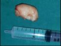
A 40 years old lady presented to our OPD
with mild discomfort in swallowing, nasal
block, left ear block on & off, hyponasal
voice for the past 6 months. On examination
a smooth mass was seen in the oropharynx
hanging from above . On Diagnostic Nasal
Endoscopy, the mass was seen to be rising
from left eustachian cushion obstructing the
left side of the choana completely. The same
mass seen partially occluding the right side
as well . No attachment to any other walls of
the nasopharynx was made out. routine investigation
were done including CT Paranasal
Sinuses and Neck. It showed the mass
extending from the nasopharynx upto the
oropharynx 6/4 cm Housefield Units ‐32 to
17. Under local anaesthesia, endoscope assisted
biopsy was taken and sent for HPE.
Under GA, endoscopic assisted excision of
the mass doneusing Radio‐Frequency Ablator
with haemostasis . Post operative period
was uneventful
Full Text:
PDFReferences
Lipoma in Fossa of Rossenmuller June J
Laryngol Otol June 2000 KalanA,Ahmed Shuaib
A,Tariq M 114(6);465‐6
Nasopharyngeal Lipoma ‐A rare clinicopathological
entity Fagan JJ 1,Learmonth
G ,Garbon,Bower R M,J Laryngol Otol 1996
Mar ;110(3):275‐6
Retronasal lipoma‐A Case Report Otolaryngol
Head &Neck;Surgery.1989 Mar;100(3)248‐
Pitts D B1,Hilsinger R L Jr
The Interesting Case‐Case no 67 Lohnstein P
V,Maier W,Birkenhager R,Schipper J Laryngorhinootogie
Jan;84(1):51‐3
LIpoma a Rare Tumourof Nasopharynx Indian
J Cancer 1997 Dec;34(4):177‐8
HugeRetropharyngeal Lipoma Causing Obstructive
Sleep Apnoea:a case report Namyslowski
G1,Scierski W,Misiolek M,Urbaniec
N,Lange D Rur Arch Otorhinolaryngol 2006 Aug
(8):738‐40.Epub 2006 May 4
Refbacks
- There are currently no refbacks.

This work is licensed under a Creative Commons Attribution-NoDerivatives 4.0 International License.
An Initiative of The Tamil Nadu Dr MGR Medical University