 University Journal of Surgery and Surgical Specialities
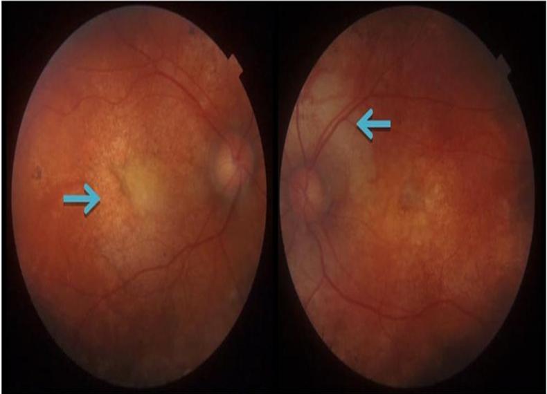
CHOROIDAL OSTEOMA – A CASE SERIES
Abstract
We report a case series of choroidal osteoma, a rare benign tumor of the eye. The diagnosis can be arrived with the help of multimodal imaging techniques of Optical Coherence Tomography (OCT) B-scan and Fundus Fluorescein Angiography. In this case series we can see typical fundus findings of yellow orange lesion commonly in juxtapapillary area and hyper reflective lines with posterior delineation in OCT and calcification with after shadowing in Bscan. Tumor can be associated with choroidal neovascular membrane (CNVM) threatening vision. In such cases intravitreal bevacizumab were helpful in restoring vision. A serial follow up of these patients is required to assess CNVM and decalcification of the osteoma.
Full Text:
PDFReferences
Kadrmas EF et al. Choroidal osteoma. Int Ophthalmol Clin. 1997;37(4):171–82
Aylward et al. A long-term follow-up of choroidal osteoma. Arch Ophthalmol. 1998 Oct;116(10):1337–41.
Sisk RA et al. Fundus autofluorescence findings of choroidal osteoma. Retina (Philadelphia, Pa). 2013 Jan;33(1):97–104.
Shields CL et al. Choroidal osteoma shows bone lamella and vascular channels on enhanced depth imaging optical coherence tomography in 15 eyes. Retina (Philadelphia, Pa). 2015 Apr;35(4):750–7.
Hussain R et al. Real-time in vivo micromorphology and histopathology of choroidal osteoma using enhanced depth imaging. Indian J Ophthalmol. 2015 May;63(5):453–5.
Refbacks
- There are currently no refbacks.

This work is licensed under a Creative Commons Attribution-NoDerivatives 4.0 International License.
An Initiative of The Tamil Nadu Dr MGR Medical University