 University Journal of Surgery and Surgical Specialities
A case of large non metallic intraorbital extraocular foreign body
Abstract
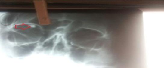
A 36 year old male presented with history of Road traffic accident to casualty, on presentation patient had vision of Hand movements in Right eye on examination patient had entry wound in lid, cornea edema with Descemet membrane folds, fundus examination could not be done due to hazy media . CT orbit revealed a large Intraorbital extraocular hyperdense foreign body, B Scan showed a large foreign body at 12 o' to 1 o' clock position with after shadowing . Foreign body was removed intoto under Local anaesthesia through anterior orbitotomy which measured 1x1 cm and was non metallic in nature. Patient has a vision of 6 by 36 post operatively and there were no signs infection This case is being presented as the foreign body was non metallic in nature which is large in size and it was removed in toto and in time which decreases infection rate and has better visual prognosis.
Full Text:
PDFReferences
Fulcher TP, McNab AA, Sullivan TJ. Clinical features and management of intraorbital foreign bodies.Ophthalmology. 2002;109:494–500.
Abdullah Al-Mujaini, Rana Al-Senawi, Anuradha Ganesh, etall, Sultan Qaboos Univ Med Jv.8(1); 2008 Mar
Refbacks
- There are currently no refbacks.

This work is licensed under a Creative Commons Attribution-NoDerivatives 4.0 International License.
An Initiative of The Tamil Nadu Dr MGR Medical University