Prevalance of Dermatophytes in Tertiary Care Hospital
Abstract
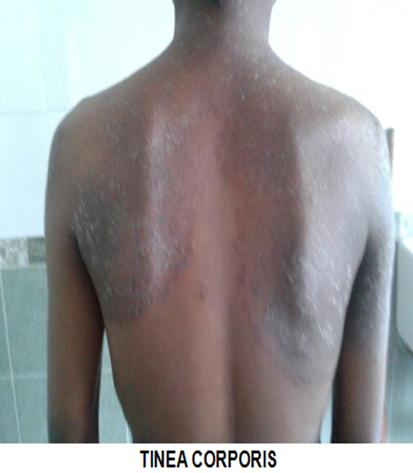
Introduction: Dermatophytoses are the infections of keratinized tissues such as the epidermis, nail and hair caused by a group of closely associated filamentous fungi called as Dermatophytes
Aim & Objectives: 1.To isolate the dermatophytes causing infection of skin, hair and nail.2.To identify and speciate the isolates of dermatophytes.3.To determine the antifungal susceptibility of the isolates.
Materials & Methods: The cross sectional study about dermatophytosis comprised of 50 patients (n=50) attending the dermatology clinic and was carried out in department of microbiology in Government Kilpauk Medical College and Hospital, Chennai for a period of 3 months (September to November 2017) to isolate and speciate the dermatophytes and to determine its antifungal susceptibility pattern.
Results: Out of 50 clinical samples, 32(64.%) were skin scrapings, 5(10%) were hair samples and 13(26%) were nail samples.Out of 50 isolates 15(30%) isolates were culture positive and 35(70%) were culture negative for dermatophytes. Trichophytonwas the most common genus in culture isolates. 9 Trichophytonrubrum were isolated 5Trichophytonmentagrophyteswere isolated and 1 Trichophytontonsuransisolated. The MIC range, MIC 50 and MIC 90 for the drug fluconazole was found to be 0.015-0.25µg/ml, 2 µg/ml and 8 µg/ml respectively. The MIC range for terbinafine by microbroth dilution method was 0.015µg/ml- 0.25 µg/ml and MIC 50 and MIC 90 was found to be 0.5 µg/ml and 2 µg/ml. Only one Trichophytonmentagrophytes shows higher MIC values (MIC 50 was 2 µg/ml) and no higher MIC inTrichophytonrubrum
Conclusion: :Specific identification of the dermatophytic species and timely institution of appropriate antifungal therapy based on the prevailing sensitivity pattern could be of immense value. The increased incidence and availability of various new drugs for dermatophytic infection in the last two decades emphasis a reference susceptibility testing method ,which aids the clinician to select the appropriate drugs for the management of dermatophytic infection.
Keywords: Dermatophytes, KOH mount, Fungal culture, Antifungal susceptibility testingFull Text:
PDFReferences
Peerapur B V,InumdarAC, PushpaPV, rikanthB, Clinicomycological Study of Dermatophytosis in Bijapur,Indian Journal of Medical Microbiology;(2004) 22 (4) : 273-274.
Topley and Wilsons, Microbiology and Microbial Infections Vol4. 10th edition (220-244).
Suman Singh, P.M.Beena, Profile of dermatophyte infections in Baroda, Indian DermatolVenereolLeprol July-August 2003 Vol 69 Issue 4.
Clarissa J.Lyngdoh, W.ValrieLyngdoh, Basabdatta Choudhury, KalkambeA.Sangma, IshaniBore, AnnieB.Khyrlem, Clinico-Mycological profile of Dermatophytosis in Meghalaya, Madanal Journal of Medical and Public Health | Oct-Dec 2013 | vol 3 Issue 4.
U.S.Agarwal, Jitendara Saran, Puneet Agarwal, Clinico-mycological study of dermatophytes in a tertiary care centre in northwest India,Indian J DermatolVenereolLeprol 2014;80 :194.
Smita Sarma, AkBorthakur, A clinico-epidermiological study of dermatophytoses in Northeast india.Indian J dermatolVenereolLeprol | November- December 2007 | Vol 73 | Issue 6.Page : 427-428
Surendaran KAK, Ramesh M Bhat, Rekha Boloor, Nanadakishore B, Sukumar D, A Clinical and Mycological Study of Dermatophytic Infections,Indian Journal of Dermatology 2014;59(3).
ArunaVyas,NazneenPathan, Rajini Sharma, LeelaVyas, A Clinicomycological Study Of Cutaneous Mycoses in Sawai Man Singh Hospital Of Jaipur, North India, Annals of Medical and Health Sciences Research | Oct-Dec 2013 | Vol 3 | Issue 4.
P Veer, NSPatwardhan, AS Damle; Study of Onchomycosis: Prevailing Fungi and Pattern of Infection, Indian Journal of Medical Microbiology(2007)25(1):53-6.
R kaur, BKashyap, P Bhalla, Onychomycosis-Epidmiology Diagnosis and Management, Indian Journal of Medical Microbiology (2008) 26(2):108-16.
Venkatesan G, Singh R, Murugessan AG, Janaki C, Shankar GS. Trichophytonrubrum-the predominant etiological agent inhumandermatophytoses in Chennai, India. Afr J Microbiol Res 2007;1:9-12.
P Kannan, C Janaki, GS Selvi, Prevalence of Dermatophytes and Other Fungal Agents Isolated From Clinical Samples, Indian Journal of Microbiology, (2006) 24 (3):212-5.
Bindu .V, Clinico-mycological study of dermatophytosis in calicut. Indian J DermatolVenerolLeprol 2002:68:259-261.
SanjeevSahai,Devesh Mishra,Change in spectrum of dermatophyes isolated from superficial mycoses cases: First report from Central India,Indian Journal of Dermatology, Venereology and Leprology | May-June 2011 | Vol 77 | Issue 3
Mohrenschlager M, SeidlHP, Ring J, et al. Pediatrictineacapitis: recognition and management .Am J clinDermatol 6: 203-213,2005.
National Committee for Clinical Laboratory Standards,2002.Reference method for broth dilution antifungal; Susccptibility testing of filamentous fungi. Approved Standard M38-A.National Committee for Clinical Laboratory Standards. Waync,Pa.
Establishing a Method of Inoculum Preparation for Susceptibility testing of Trichophytonrubrum and T.mentagrohytes.D a Santos, M.E.S Barros and J.S HamdanJCM,Jan 2006,p98-100.
VanathiSabtharishi, Rathikakatragadda, Thyagarajanravinder, a study of the antifungal susceptibility pattern of dermatophytes isolated in a tertiary care hospital. Int .J. bioassay vol6(5) 2017
Suganthi M et al, Antifungal agents and their action against dermatophytes: curious to know the facts, Journal of innovations in pharmaceuticals and biological sciences . e. ISSN: 2349- 2759
VK Bhatia, PC Sharma, Determination of minimum inhibitory concentrations of itraconazole, terbinafine and ketoconazole against dermatophyte species by broth microdilution method: 2015vol.33(4),533-537
Refbacks
- There are currently no refbacks.

This work is licensed under a Creative Commons Attribution-NoDerivatives 4.0 International License.
An initiative of The Tamil Nadu Dr M.G.R. Medical University