Anatomical study of the Triangle of Brocq and Mouchet with its clinical implications
Abstract
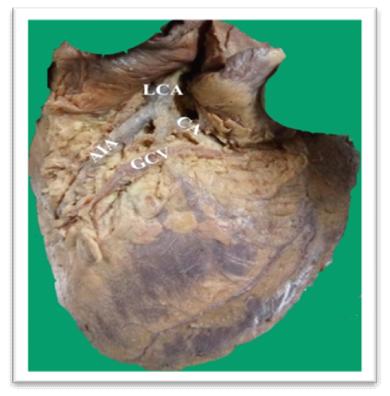
The triangle of Brocq and Mouchet is situated on
the left side of the sternocostal surface and is formed by the
crossing between the anterior interventricular artery,
the circumflex branch of the left coronary artery and the
great cardiac vein. In the present study, 43 cadaveric heart
specimens from the Department of Anatomy, Christian
Medical College were dissected and the topography of the
triangle based on the position of the vessels were classified
and tabulated. This study may provide useful information for
interventional cardiac procedures.
Full Text:
PDFReferences
Sousa-Rodrigues D, Fernando C, Soares de Alcântara F,
Silva V da, Nixon W, Soares de Alcântara F, et al. TRÍGONO
ARTERIO-VENOSO DEL CORAZÓN (DE BROCQ &
MOUCHET). Int J Morphol. 2004 Dec;22(4):291–6.
Pejkovic B, Bogdanovic D. The great cardiac vein. Surg
Radiol Anat SRA. 1992;14(1):23–8.
Aoki J, Rodríguez-Granillo GA, Serruys PW,
Aoki J, Rodríguez-Granillo GA, Serruys PW. Emergent
Strategies in Interventional Cardiology. Rev Esp Cardiol. 2005
Jan 8;58(08):962–73.
Bharathi SB. An Anatomical Study of Triangle of Brocq &
Mouchet in Human Cadaveric Heart & Its Clinical Relevance.
IOSR Journal of Dental and Medical Sciences. Jul.- Aug.
; 8(2): 12-15.
Andrade R. Triangle of Brocq and Mouchet: anatomical
study in braziliancadavers and clinical implications.
J. Morphol. Sci., 2010, vol. 27, no. 3-4, p. 127-129.
Asirvatham SJ, Stevenson WG. Editor’s Perspective:
Similia Similibus Curantur. Circ Arrhythm Electrophysiol. 2013
Dec 1;6(6):e85–6.
Refbacks
- There are currently no refbacks.

This work is licensed under a Creative Commons Attribution-NoDerivatives 4.0 International License.
An initiative of The Tamil Nadu Dr M.G.R. Medical University